What is a sentinel lymph node biopsy for melanoma?
Why sentinel lymph node biopsy is offered after a melanoma diagnosis, who it is recommended for, what happens on the day, recovery and risks, and what a positive or negative result means for you.

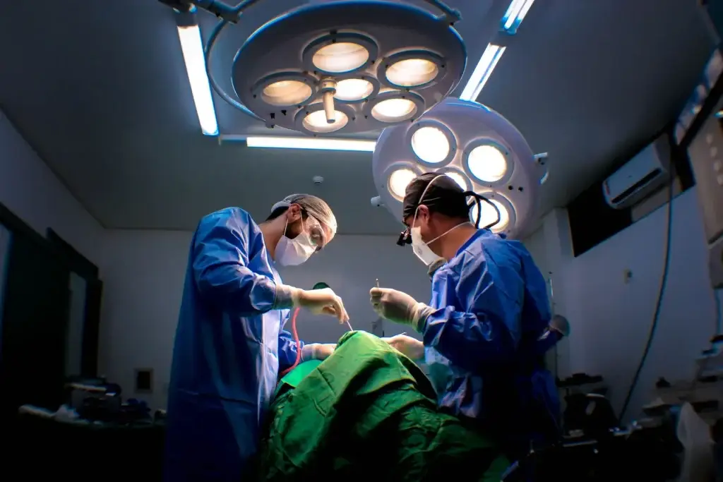
The short answer: a sentinel lymph node biopsy finds and removes the first lymph node that a melanoma would drain to, so it can be checked under the microscope. It tells us whether the melanoma has started to spread, which helps decide whether you need any further treatment. It is offered for melanomas that are thicker than about 1 mm, or slightly thinner ones with higher-risk features, and is usually done at the same time as the wide local excision, as a day case under general anaesthetic.
Sentinel node biopsy is a big part of my work. I was involved in setting up the service for melanoma at Queen Victoria Hospital in East Grinstead, and I lead the skin cancer multidisciplinary team for Kent and the local Sussex area. A melanoma diagnosis is frightening, and the weeks of tests and results can feel very long, so here are the questions patients most often ask me, answered as clearly as I can.
“A sentinel node biopsy is about information. It gives us the clearest picture of where things stand, so that every decision after it is based on fact rather than guesswork.”
Mr Siva Kumar, Consultant Plastic Surgeon
What is a sentinel lymph node?
Lymph nodes are small, bean-shaped glands that filter fluid, called lymph, from the skin and the rest of the body. Each area of skin drains through tiny lymph channels to a particular group of nodes: usually the armpit for the arm, chest and upper back, the groin for the leg and lower body, and the neck for the head.
When melanoma spreads, the first place it usually goes is the nearest group of lymph nodes. The sentinel node is the first node, or sometimes the first two or three, in that chain. If it is clear, it is very unlikely that the melanoma has spread to the other lymph nodes. If it contains melanoma cells, finding that early allows us to plan further treatment.
Who is offered a sentinel node biopsy?
It is not offered to everyone. It depends on the thickness and features of your melanoma, which your pathology report describes. The most important measurement is the Breslow thickness, how deep the melanoma has grown, in millimetres. In line with NICE guidance in the UK, a sentinel node biopsy is:
- not usually needed for thin melanomas (stage IA), where the chance of spread is very low
- considered for melanomas 0.8 mm to 1.0 mm thick with at least one higher-risk feature: ulceration, lymphovascular invasion, or a mitotic rate of 2 or more
- considered for all melanomas thicker than 1.0 mm
Other things also matter: your general health, where the melanoma is, and what you would like to know. For example, melanomas on the head and neck can drain in less predictable directions, which makes the operation more complex. I will explain the benefits and the risks for your situation, and we decide together. It is completely reasonable to choose not to have it.
Why have a sentinel node biopsy?
A sentinel node biopsy is mainly a staging test. It does not in itself cure melanoma, but it is the most accurate way to find out whether microscopic melanoma cells have reached the lymph nodes, long before they could be felt or seen on a scan. That information:
- gives the most accurate picture of your outlook
- decides whether you might benefit from drug treatment to reduce the risk of the melanoma coming back
- shapes how often you are followed up and whether you need scans
- can open the door to clinical trials
How is the sentinel node found?
Before surgery, a small amount of radioactive tracer is injected into the skin near the melanoma scar. It travels through the lymph channels to the sentinel node, and a scan, called lymphoscintigraphy, shows where it is. This is usually done on the morning of surgery or the day before.
During the operation, a blue dye is also injected around the scar. Using a handheld probe, which picks up the tracer, and the blue colour, I can find the exact node and remove it through a small incision, usually a few centimetres long. The amount of radioactivity is very small, less than many routine X-rays, and it fades quickly.
What happens on the day
The sentinel node biopsy is usually done alongside your wide local excision, which removes a margin of skin around the original melanoma scar to reduce the chance of it coming back locally. The margin is usually 1 cm for stage I melanoma and 2 cm for stage II. The wound is closed directly, or with a local flap or skin graft if needed.
You have a general anaesthetic, and most people go home the same day. The whole operation usually takes one to two hours, depending on where the melanoma and the sentinel node are.
Sentinel lymph node biopsy recovery
- Pain: most people need only simple painkillers for a few days. The node site often feels sorer than the excision site.
- Dressings: keep them dry for the first few days.
- Stitches: removed or dissolve within about one to two weeks.
- Activity: avoid strenuous activity, heavy lifting and stretching the area for the first couple of weeks. Gentle walking is encouraged.
- Work: most people with desk jobs return within one to two weeks, depending on where the surgery was.
- Driving: once you can move comfortably and do an emergency stop, often after a week or so.
- Blue colour: the dye can turn your skin or urine a blue or green colour for a while. This is harmless and temporary, although a faint blue stain near the scar can take some months to fade.
What are the risks?
The most common problem is a collection of fluid where the node was removed, called a seroma. It sometimes needs draining in clinic with a needle. Wound infection, bleeding and numbness around the scar can happen. Swelling of the limb, called lymphoedema, is uncommon after sentinel node biopsy alone, and slightly more likely after surgery in the groin than the armpit, but it is something I will discuss with you. Very rarely, people have an allergic reaction to the blue dye, which the anaesthetist is prepared for.
No test is perfect. Occasionally, in a small number of people, the sentinel node is clear but melanoma later appears in another node in the same area. That is one reason follow-up continues afterwards.
Getting the result
The node is examined in detail in the laboratory, and results usually take two to three weeks. Your case is reviewed by the specialist skin cancer multidisciplinary team, and I will go through the results with you in person. I know this wait is hard.
If the sentinel node is clear (negative)
This is the result for most people, roughly four in five. It means it is very unlikely that the melanoma has spread to your lymph nodes. You move on to regular follow-up and skin checks, and the frequency depends on the stage of your melanoma.
If the sentinel node is positive
If it contains melanoma cells, your melanoma is classed as stage III. That sounds alarming, but many people in this position do very well. Your case is discussed at the specialist skin cancer MDT, and you will usually be offered:
- scans, such as a CT or MRI, to check the rest of the body
- regular ultrasound scans of the lymph nodes in that area, rather than an operation to remove them all, which large studies have shown is not usually necessary
- adjuvant treatment, such as immunotherapy or targeted drugs, which can lower the risk of the melanoma coming back
“Melanoma treatment has changed beyond recognition in the last decade. A positive sentinel node today means very different things from what it meant even ten years ago.”
Mr Siva Kumar, Consultant Plastic Surgeon
Looking after yourself afterwards
Whatever the result, learning to check your own skin and lymph nodes is one of the most useful things you can do between appointments. I have written a step-by-step guide on how to check your lymph nodes after melanoma, and another on checking your moles. Sun protection, and seeing your team promptly about anything new, matter for life.
Can I have a sentinel node biopsy privately?
Yes. Most private insurers cover melanoma treatment, including sentinel node biopsy, and you can also self-fund. Being seen privately can mean faster appointments and seeing the same consultant throughout, while your care is still reviewed by the specialist skin cancer team.
Related question: Private skin cancer surgery: why would you choose it?
Questions I’m often asked
Is a sentinel lymph node biopsy necessary for melanoma?
It is optional. It does not cure melanoma on its own, but it gives the most accurate information about stage, which guides treatment decisions. Some people decide the information is not worth an operation for them, and that is a valid choice.
Does a sentinel node biopsy hurt?
You are asleep for the operation. Afterwards, most people describe soreness and stiffness rather than severe pain, settling over one to two weeks.
How long does a sentinel lymph node biopsy take?
The scan beforehand takes an hour or two. The operation, combined with the wide local excision, usually takes one to two hours.
Can it be done if I am pregnant?
The options, including delaying the biopsy until after pregnancy, are discussed individually with the specialist team.
Next steps
If you have recently been diagnosed with melanoma and want to talk through your options, you are welcome to book a consultation. There is more detail on my melanoma treatment page, and the full UK guidance is published by NICE (NG14).

Mr Siva Kumar Veeramani
MBBS, BMed.Sci (Hons), MRCS, MBA, FRCS (Plast.). Consultant Plastic, Reconstructive & Cosmetic Surgeon practising across London, Kent, Surrey and Sussex. GMC 6114710.
About Mr Siva Kumar →